Although they have been used for decades, CT scans are a relatively new procedure when compared with X-rays. For that reason, if one of our doctors here in Southfield has advised you to have images taken, you may have wondered what the differences are between an X-ray and a CT scan.
Both are excellent imaging techniques and can be used for similar parts of the body, after all, so why is one chosen over the other?
Understanding how the different technologies work may help.
Facts about X-Rays
Let’s start with a fun fact: Do you know what the X in X-ray stands for? We’ll give you a hint: What does the “X” mean in an algebra equation?
You got it! Same answer.
The X literally stands for “unknown.” When German physicist Wilhelm Roentgen discovered this form of radiation in 1895, he referred to it as “X” because it was still that mysterious to him.
And we’re sure you can guess that the “ray” refers to radiation.
The phrase “unknown radiation” might not exude much confidence, but fortunately science has come a long way since 1895.
We know much more about it now. In fact, here’s how it works.
An X-ray is a form of high-energy electromagnetic radiation that can pass through most objects, including the body. The rays travel through the body and strikes an X-ray detector (such as a radiographic film or a digital X-ray detector) on the other side of the patient.
The images that are created are due to the calcium in the body. The calcium is a denser material than most other tissues and therefore can stop some of the X-rays. As a result, an image is formed that represents the “shadows” of objects inside the body.
You will notice that you can see the bones rather vividly (even though it’s in black and white), but the other tissues are shadowy outlines.
Although they can be used to view internal organs, X-rays are most often used if the doctor needs to examine bone, such as in the case of a fracture.
X-rays are 2-dimensional images.
In Michigan, X-rays cost about $100 to $900 on average.

An X-ray is a 2-dimensional image that is most often used to analyze bone.
Facts about CT Scans
Now that you have a general idea about how X-rays capture images, it may be easier to understand how CT scans work.
CT scans are essentially a series of X-rays that are stacked on top of one another to create a 3-dimensional image. Also known as CAT scans, CT scans use computer technology to create cross-sectional 2D “slices” of that area. These slices are then digitally stacked together to create this 3D image.
As a result, instead of one, flat image in an X-ray, you can see a fuller shape with a CT scan. The image quality is also better.
Depending on the area being imaged, the cost of a CT scan in Michigan ranges from less than $250 to nearly $4,000.

A CT scan uses multiple layers of X-rays to form a 3-dimensional image.
The Differences Between X-Rays and CT Scans
On a basic level, one of the main differences between X-rays and CT scans is the cost. Many providers will start with an X-ray to determine if that is sufficient for diagnosis. If it’s not, they may recommend a more detailed, comprehensive image through a computerized tomography scan.
Another important difference between the two imaging techniques is the process and duration. An X-ray typically can be taken on the spot and only takes a few minutes for each image. The patient would lie on a table or sit in a chair (such as for a simple X-ray of a hand), and the patient may be asked to move positions to enable the radiologist to take the necessary pictures.
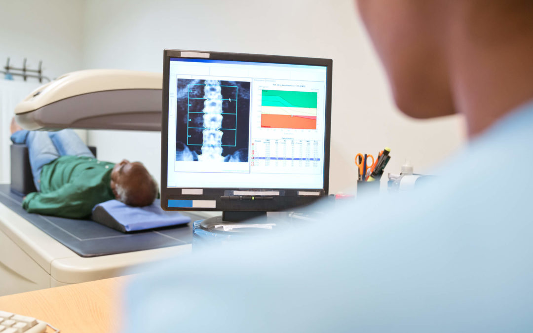
In a CT scan, the patient would lie on a table, and the radiologist or other medical professional may help position the patient’s body optimally for the best images and the patient’s comfort. Afterward, a doughnut-like structure called a gantry would circle around the patient as images are taken. The entire machine looks a little like a tunnel, and it is usually not fully enclosed.
Another difference between X-rays and CT scans is the duration. X-rays typically don’t take more than 15 minutes, even if multiple images are taken. A CT scan takes a minimum of 15 minutes but closer to a half hour.
A fourth, significant difference is the need to sometimes use a dye, referred to as contrast, when CT scans are taken. The dye would be injected into the body prior to the procedure. If the dye is required, it may increase the cost of the imaging technique, and some patients have an adverse reaction to the dye.
If you want to know more about the differences between X-rays and CT scans and the out-of-pocket costs, contact our office here in Southfield, Michigan, or make an appointment to see one of our doctors.