

Echocardiogram cardiac ultrasound testing is available in our Southfield, Michigan practice.
An echocardiogram uses high-frequency sound waves to produce images of your heart, including the chambers, valves, walls, and blood vessels. This commonly used test will allow our physicians to literally see your heart beating and pumping blood. The doctor can use the test to identify heart disease and prevent heart failure and heart attack before it happens.
Dr. Feinstein may recommend an echocardiogram cardiac ultrasound if she suspects problems with the valves or chambers of your heart. You may also be advised to take the heart test if you are experiencing symptoms consistent with heart disease, such as shortness of breath or chest pain.

What Happens During an Echocardiogram Cardiac Ultrasound
During an echocardiogram cardiac ultrasound, a probe called a transducer is passed over your chest as you lie on your back. The tool produces sound waves that bounce off your heart and “echo” back to the probe, which are changed into pictures that the doctor can view on a monitor.
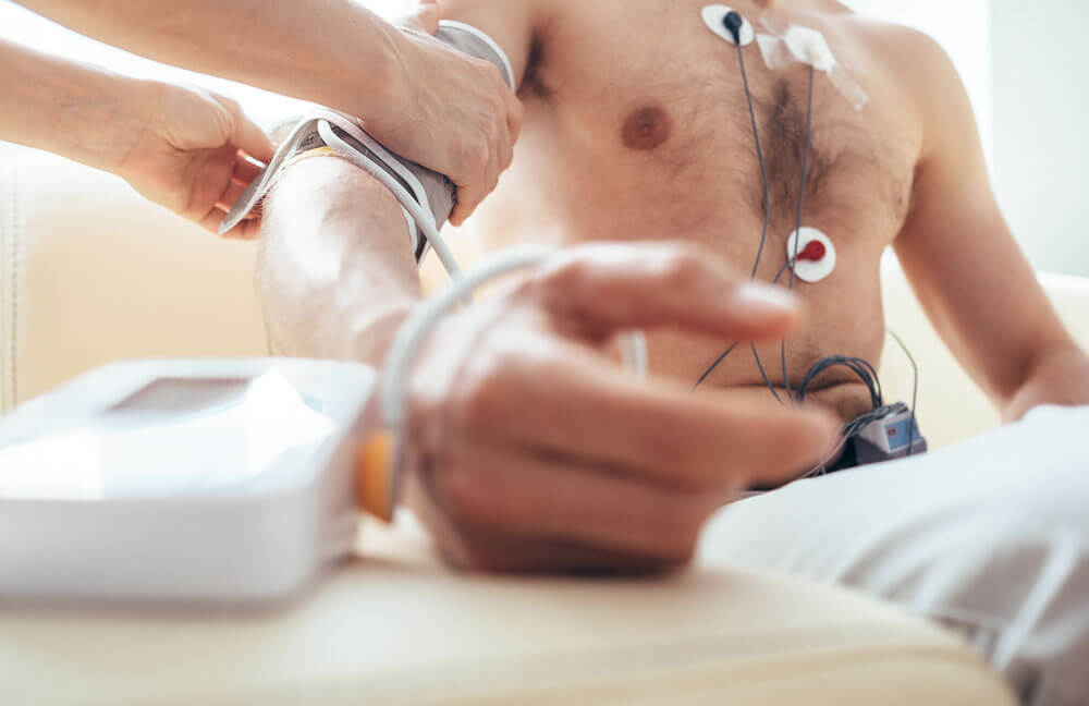
To start the test, you will be asked to lie on your back, and small metal disks will be placed on your chest. These disks have wires that connect to a machine that tracks your heart rate.
The doctor or technician may place a gel on your chest to help sound waves pass through your skin, and you may be asked to move or hold your breath briefly for better pictures.
The room may be dark so that the technician can see the images better.
The doctor will discuss findings and echocardiogram results with you. If any concerns are detected, we will work with you to devise a heart treatment plan that works for you.
For more information about echocardiogram testing, message our Southfield medical office online or call for an appointment.